Services
Detection Of Early Gastric & Esophageal Cancers With NBI Mode
Detection of early gastric and esophageal cancers with Narrow Band Imaging (NBI) mode involves the use of specialized endoscopic technology to enhance visualization of the mucosal surface and microvasculature in the gastrointestinal tract. Here are the details:
Principle of NBI Mode
- NBI utilizes narrow-bandwidth light filters to highlight specific wavelengths of light, primarily in the blue and green spectrum.
- By narrowing the bandwidth of light, NBI enhances the visualization of superficial tissue structures and microvascular patterns within the mucosa.
- This technique provides better contrast and delineation of abnormalities compared to conventional white-light endoscopy, making it particularly useful for detecting subtle mucosal changes associated with early-stage cancers.
Endoscopic Procedure
- During an endoscopic examination, the endoscopist switches the endoscope to NBI mode, activating the specialized light filters.
- The NBI mode is used to carefully inspect the mucosal surface of the stomach and esophagus for any abnormalities, such as irregularities in color, vascular patterns, or surface morphology.
- The endoscopist systematically examines the entire mucosa, paying close attention to areas of concern based on patient history, symptoms, or previous findings.
Identification of Suspicious Lesions
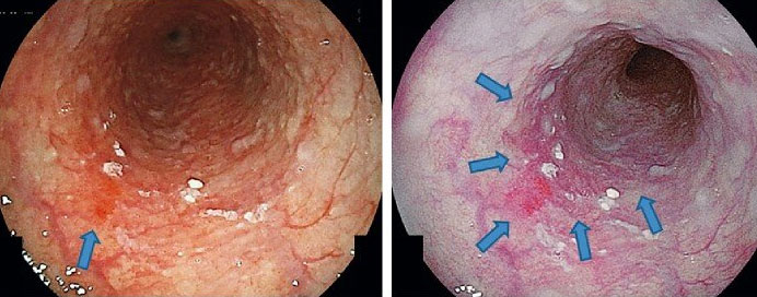
- In early gastric and esophageal cancers, subtle changes in mucosal appearance and vascular patterns may indicate the presence of neoplastic lesions.
- With NBI mode, abnormal vascular patterns associated with early-stage cancers, such as irregular vessels or avascular areas (gaps in the normal vascular network), are more readily identified.
- Suspicious lesions, including flat or depressed lesions, nodules, or areas of irregular mucosal surface, are carefully evaluated under NBI mode for further assessment.
Biopsy and Histological Confirmation
- If suspicious lesions are identified, targeted biopsies are performed to obtain tissue samples for histological examination.
- Biopsies are taken from the abnormal areas visualized under NBI mode to confirm the presence of neoplastic changes and determine the histological grade and extent of the lesion.
- Histological confirmation of early gastric and esophageal cancers allows for accurate diagnosis and staging, guiding subsequent treatment decisions.
Clinical Applications and Benefits
- NBI mode is particularly valuable for the surveillance and early detection of gastric and esophageal cancers, especially in high-risk populations.
- Early detection of neoplastic lesions with NBI can lead to timely intervention, improved treatment outcomes, and potentially better long-term survival for patients.
- Additionally, NBI mode can aid in the differentiation of neoplastic lesions from benign conditions or inflammatory changes, reducing the need for unnecessary biopsies.